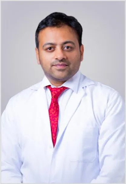
Knee Arthroscopy Surgeon in Bangalore | Advanced Keyhole Knee Surgery
What is Knee Arthroscopy?
Knee arthroscopy is a sophisticated, minimally invasive surgical procedure that allows a specialist surgeon to visualize the knee joint using a high-definition camera. During the procedure, your Orthopedic Surgeon in Bangalore inserts an arthroscope, a slender, fiber-optic instrument through tiny, keyhole incisions to diagnose and treat various knee injuries precisely with minimal tissue damage.

Excellence in Healthcare
State-of-the-art facilities & specialist doctors
Who Requires Knee Arthroscopy?
If persistent knee pain fails to improve with rest, physical therapy, or non-surgical interventions, a healthcare provider may recommend arthroscopic knee surgery Bangalore.
This procedure is essential for diagnosing injuries to the ligaments and cartilage that may not be fully visible on an MRI. It is particularly common among athletes in Bangalore who participate in high-impact sports involving jumping, running, or sudden pivoting. While arthroscopy is highly effective for mechanical issues, it is generally not the primary treatment for advanced osteoarthritis.
Why Does Your Surgeon Perform Knee Arthroscopy?
A specialized surgeon performs an arthroscopy to achieve three goals: accurate diagnosis, targeted repair, and tissue removal in Bangalore. By utilizing real-time imaging, the doctor can:
Diagnose: Identify the
exact source of mechanical pain or locking.
Repair: Reconstruct
torn ligaments (ACL/ PCL
) or suture damaged tendons using specialized surgical equipment.
Remove: Resect inflamed
synovial tissue or trim damaged cartilage that causes friction within the joint.
Symptoms That Indicate a Knee Problem
When a knee joint experiences structural damage or inflammation, it typically sends distinct warning signs. Recognizing these indicators early can prevent minor issues from developing into chronic joint damage.
- Mechanical Locking or Catching: A physical sensation where the knee suddenly gets "stuck" at a specific angle and refuses to straighten or bend, often caused by a torn meniscus flap or a loose bone/cartilage fragment blocking the joint path.
- Persistent Deep Joint Pain: A dull, aching, or sharp localized pain within the knee that intensifies during weight-bearing activities, twisting movements, or climbing stairs, and fails to subside with rest or over-the-counter anti-inflammatory medications.
- Recurrent Swelling (Effusion): Fluid buildup inside the knee joint capsule that occurs within hours of activity. It often presents as a visibly puffy knee that feels warm to the touch or tight when bending.
- Instability or "Giving Way": A sudden feeling that the knee is unstable, buckling, or shifting out of place during routine walking, pivoting, or step-climbing, which frequently points to a ligament tear such as an ACL or PCL injury.
- Audible Popping or Grinding: A distinct popping sound or clicking sensation felt inside the joint during motion, frequently accompanied by localized pain or a feeling of friction.
When to See an Orthopaedic Surgeon
Not all knee pain requires immediate surgical attention, but certain red flags mean you should book a clinical evaluation with an orthopaedic specialist. You should consult a surgeon if you experience:
- Inability to Bear Weight: You cannot stand on the affected leg or take more than a couple of steps without severe pain or buckling.
- Failed Conservative Treatment: Your knee pain, swelling, or stiffness persists after 4 to 6 weeks of targeted rest, activity modification, icing, and structured physical therapy.
- Functional Limitations: Your knee issues interfere with daily activities, work productivity, sleep patterns, or prevent you from participating in sports or fitness routines.
- Visible Joint Deformity: The knee looks noticeably misaligned, structural changes are visible, or there is localized, extreme redness and heat surrounding the joint.
- Signs of Infection or Nerve Compression: Unexplained fever combined with knee pain, radiating numbness, or a "pins and needles" sensation traveling down your lower leg or foot.
How Your Condition is Diagnosed
An accurate diagnosis is the cornerstone of effective treatment. Dr. Pradyumna uses a comprehensive, step-by-step diagnostic protocol to pinpoint the exact structural cause of your knee symptoms:
- Clinical History and Evaluation: A detailed discussion about the onset of your pain, injury mechanisms, lifestyle demands, and prior treatments, followed by a careful physical exam.
- Specialized Physical Provocative Tests: The surgeon performs manual joint stability assessments such as the Lachman test (for ACL integrity) or the McMurray test (to check for meniscus tears) to evaluate ligament laxity and cartilage tenderness.
- Digital X-rays: Performed to assess bone alignment, identify potential fractures or bone chips, and rule out severe joint space narrowing caused by advanced osteoarthritis.
- High-Resolution MRI (Magnetic Resonance Imaging): The primary non-invasive tool used to visualize soft tissue structures in detail. It clearly maps out meniscus tears, ligament tears, tendon injuries, and cartilage defects.
- Diagnostic Arthroscopy (The Gold Standard): If non-invasive imaging is inconclusive but mechanical symptoms persist, a diagnostic keyhole arthroscopy allows the surgeon to directly visualize the living tissues inside the joint via a high-definition camera. This step often transitions immediately into a therapeutic repair during the same session.
Expanded Conditions Treated
Arthroscopic surgery treats a broad spectrum of structural, mechanical, and inflammatory knee problems:
Meniscal Lesions:
- Meniscus Tears (Bucket-handle, Radial, Complex): Trimming down non-viable, fraying pieces (partial meniscectomy) or preserving and suturing healthy cartilage tissue back together (meniscus repair).
- Discoid Meniscus: Contouring a congenitally thick, abnormally shaped meniscus into a functional, crescent shape to eliminate clicking and pain.
Ligament Injuries:
- Anterior Cruciate Ligament (ACL) Tears: Reconstructing a ruptured ACL using an autograft (usually hamstring or patellar tendon) to restore complete pivoting stability.
- Posterior Cruciate Ligament (PCL) Tears: Repairing or reconstructing the central stabilizing ligament to stop abnormal backward sliding of the shinbone.
- Multi-Ligament Instability: Addressing combined injuries (e.g., ACL plus MCL or LCL) to rebuild comprehensive structural integrity.
Cartilage and Bone Defects:
- Osteochondral Lesions & Cartilage Defects: Utilizing advanced techniques like microfracture (drilling tiny holes to stimulate fibrocartilage growth) or chondroplasty to smooth down roughened, damaged joint surfaces.
- Loose Bodies: Safely locating and extracting floating fragments of bone or cartilage that cause painful mechanical locking or grinding.
Synovial and Patellar Pathologies:
- Chronic Synovitis: Removing inflamed, thickened joint lining tissue caused by repeated trauma, gout, or early-stage inflammatory arthritis.
- Patellar Maltracking: Performing a lateral retinacular release or patellar realignment to guide the kneecap smoothly along its track and relieve anterior knee pain.
- Popliteal (Baker's) Cysts: Addressing the internal joint pathology (like a meniscus tear) that causes fluid buildup, resolving the painful cyst behind the knee.
Detailed Recovery Timeline
Because every patient heals at a unique pace depending on the exact type of repair performed, recovery is divided into distinct, manageable milestones:
| PHASE / TIMELINE | ACTIVITY AND MOBILITY STATUS | PAIN AND SWELLING MANAGEMENT | EXPECTED MILESTONES |
|---|---|---|---|
|
Phase 1
Acute Healing
(Days 1 to 14)
|
|
|
|
|
Phase 2
Progressive Mobility
(Weeks 2 to 6)
|
|
|
|
|
Phase 3
Strengthening
(Weeks 6 to 12)
|
|
|
|
|
Phase 4
Functional Return
(Months 3 to 6+)
|
|
|
|
Physiotherapy & Rehabilitation Protocols
Surgery fixes the structural mechanical problem, but targeted physical therapy restores your movement, power, and long-term joint function. Your rehabilitation program is divided into three key pillars:
Phase-Specific Therapeutic Goals
- Early Phase (Focus: Range of Motion & Activation): Emphasizes passive and active-assisted range-of-motion drills like heel slides and wall sits. Focuses on waking up the quadriceps muscle through isometric contractions (squeezing a rolled towel under the knee) to prevent rapid muscle wasting.
- Mid-Phase (Focus: Strength & Proprioception): Shifts toward progressive resistance exercises. Introduces balance boards and single-leg stability exercises to rebuild proprioception (your brain's awareness of your joint's position in space), which protects the knee from future re-injury.
- Late Phase (Focus: Power & Agility): Introduces plyometrics (explosive jumping and landing drills), side-shuffling, and sport-specific training tailored to your lifestyle whether that means climbing stairs easily or returning to a cricket match.
Compliance and Success Indicators
- Consistency is Key: A standard rehabilitation course runs for 4 to 12 weeks, requiring 2-3 structured clinical sessions per week combined with a daily home exercise plan.
- Long-Term Joint Protection: Rebuilding strong quadriceps and hamstrings acts as a natural shock absorber for the knee, significantly slowing down future joint wear and tear and protecting your surgical outcome for years to come.
Common Conditions Treated
As a leading Knee Specialist in Bangalore, Dr. Pradyumna utilizes arthroscopy to treat:
Soft Tissue Injuries:
Including ACL tears, Bursitis, Sprains, and Tendinitis.
Intra-articular
Fractures: Repairing bone chips or cartilage fragments.
Inflammatory
Conditions: Treating Synovitis (inflammation of the joint lining) caused by
infection or trauma.
How to Prepare for Your Procedure
Preparation is key to a smooth recovery. Ensure your surgeon has a complete list of all medications, including herbal supplements and vitamins.
- Fasting: Follow specific instructions regarding when to stop eating/drinking before surgery.
- Anesthesia: Depending on the complexity, you may receive local, regional, or general anesthesia. Local anesthesia numbs only the knee, while regional anesthesia (like a spinal block) numbs the lower half of your body.
The Procedure: What Happens Inside the OT?
During the surgery, your leg is prepared in a sterile environment. The surgeon irrigates the knee with a saline solution to expand the joint and clear the view.
Incision: Small portals
(incisions) are made.
Visualization: The
arthroscope sends high-resolution images to a monitor.
Correction: Specialized
instruments are used for:
- ACL/PCL Reconstruction
- Meniscus Repair or partial meniscectomy
- Patellar (Kneecap) Realignment
- Removal of Baker's Cysts or loose bone fragments
The procedure typically lasts between 30 minutes to 2 hours. Each incision is closed with a single stitch or sterile adhesive strips.
Recovery and Post-Operative Care
Post-surgery, the focus shifts to R.I.C.E. (Rest, Ice, Compression, Elevation).
- Weeks 0-2: Focus on rest and managing swelling. Use crutches as directed.
- Weeks 2-4: Begin structured Physical Therapy. This includes range-of-motion exercises and progressive weight-bearing.
- Long-term: Advanced "cutting" and pivoting exercises help athletes return to sports safely while preventing muscle atrophy.
Advantages of the Arthroscopic Approach
Compared to traditional open surgery, knee arthroscopy offers:
- Minimal Scarring: Incisions are only a few millimeters long.
- Faster Recovery: Patients often return to daily activities much sooner.
- Less Pain: Minimal trauma to muscles and bone leads to a more comfortable recovery.
Risks and Safety
Complications are rare, occurring in approximately 1% of cases. Potential risks include blood clots, stiffness, or minor infection. Most issues are easily managed through standard post-operative protocols.
When to Contact Your Doctor
Contact Bangalore Orthopaedic Clinic immediately if you experience:
- Fever or chills (signs of infection).
- Severe pain that does not respond to medication.
- Sudden, significant swelling or warmth in the calf.